Esophageal cancer
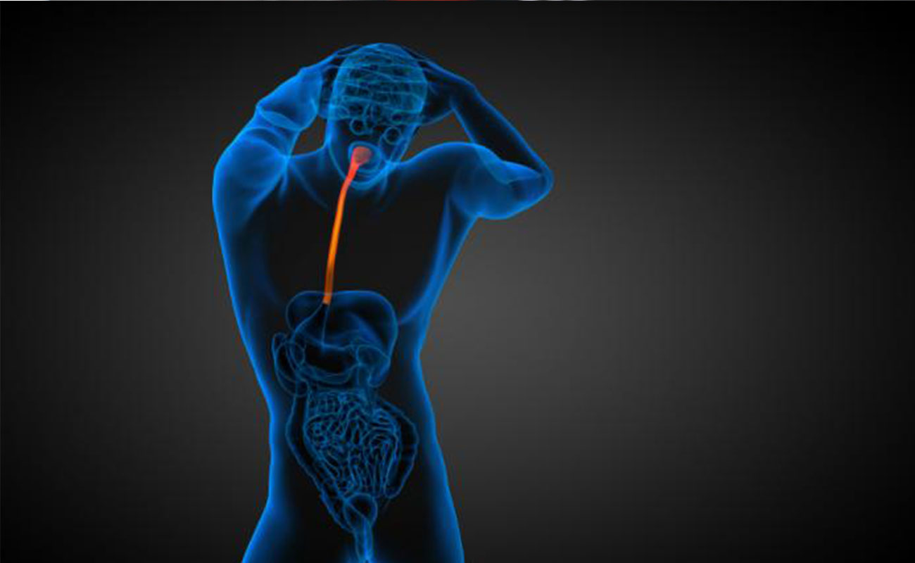
it is a disease in which malignant (cancer) cells form in the tissues of the esophagus. The esophagus is a muscular tube that moves food and liquids from the throat to the stomach.

The most common types of esophageal cancer are squamous cell carcinoma and adenocarcinoma. Squamous cell carcinoma begins in flat cells lining the esophagus. Adenocarcinoma begins in cells that make and release mucus and other fluids.
Symptoms
People with esophageal cancer may experience the following symptoms or signs. Sometimes, people with esophageal cancer do not show any of these symptoms. Or, these symptoms may be caused by a medical condition that is not cancer.
- Difficulty and pain with swallowing, particularly when eating meat, bread, or raw vegetables. As the tumor grows, it can block the pathway to the stomach. Even liquid may be painful to swallow.
- Pressure or burning in the chest
- Indigestion or heartburn
- Vomiting
- Frequent choking on food
- Unexplained weight loss
- Coughing or hoarseness
- Pain behind the breastbone or in the throat
If you are concerned about one or more of the symptoms or signs on this list, please talk with your doctor. Your doctor will ask how long and how often you’ve been experiencing the symptom(s), in addition to other questions. This is to help find out the cause of the problem, called a diagnosis.
If cancer is diagnosed, relieving symptoms remains an important part of cancer care and treatment. This may also be called symptom management, palliative care, or supportive care. Be sure to talk with your health care team about symptoms you experience, including any new symptoms or a change in symptoms.
Causues
The exact cause of oesophageal cancer is unknown, but certain things can increase the risk of it developing.
Alcohol
Drinking too much alcohol causes irritation and inflammation in the lining of the oesophagus.
If the cells in the lining of your gullet become inflamed, they're more likely to become cancerous.
Smoking
Tobacco smoke contains many harmful toxins and chemicals. These substances irritate the cells that make up the lining of the oesophagus, which increases the likelihood that they will become cancerous.
The longer you smoke, the greater your risk of developing oesophageal cancer.
Obesity
If you're overweight or obese, your risk of developing cancer of the oesophagus is higher than people of a healthy weight. The more overweight you are, the higher the risk.
This may be partly because obese people are more at risk of developing GORD and Barrett's oesophagus (see above).
Diet
Not eating enough fruit and vegetables may increase your risk of getting oesophageal cancer.
You should aim to eat at least five portions of fresh fruit and vegetables every day.
Other medical conditions
Certain rare medical conditions can also increase your chances of developing cancer of the oesophagus, including:
Achalasia – where the oesophagus loses the ability to move food along, causing vomiting and acid reflux
Paterson-Brown Kelly syndrome (also called Plummer Vinson syndrome) – a condition that can cause iron deficiency anaemia and small growths in the throat
tylosis – an inherited skin condition,
Diagnosis
Doctors use many tests to diagnose cancer and find out if it has spread to another part of the body, called metastasis. Some tests may also determine which treatments may be the most effective. For most types of cancer, a biopsy is the only way to make a definitive diagnosis of cancer. If a biopsy is not possible, the doctor may suggest other tests that will help make a diagnosis. Imaging tests may be used to find out whether the cancer has spread.
This list describes options for diagnosing this type of cancer, and not all tests listed will be used for every person. Your doctor may consider these factors when choosing a diagnostic test:
- Age and medical condition
- Type of cancer suspected
- Signs and symptoms
- Previous test results
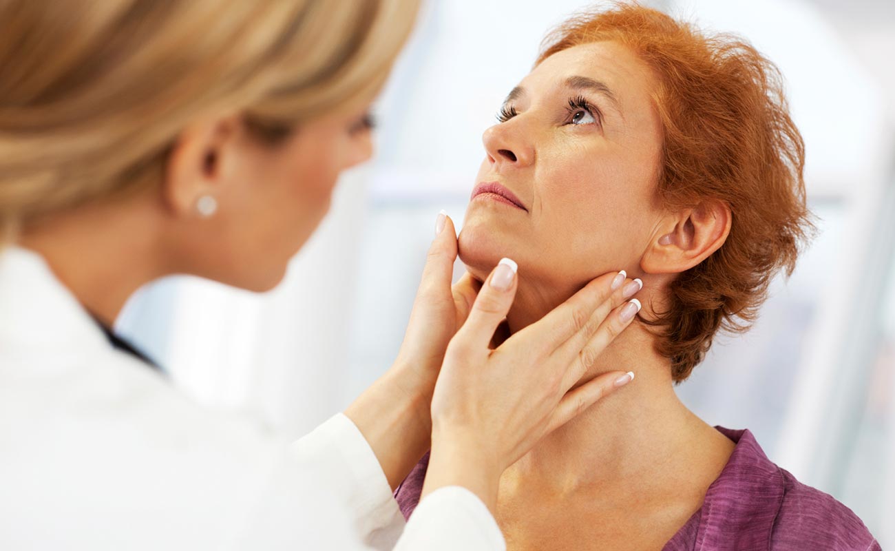
In addition to a physical examination, the following tests may be used to diagnose esophageal cancer:
- Barium swallow, also called an esophagram. The patient swallows a liquid containing barium and then a series of x-rays are taken. An x-ray is a way to take a picture of the inside of the body. Barium coats the surface of the esophagus, making a tumor or other unusual changes easier to see on the x-ray. If there is an abnormal looking area, your doctor may recommend an upper endoscopy and biopsy to find out if it is cancerous (see below).
- Upper endoscopy, also called esophagus-gastric-duodenoscopy, or EGD. An upper endoscopy allows the doctor to see the lining of the esophagus. A thin, flexible tube with a light and video camera on the end, called an endoscope, is passed down the throat and into the esophagus while the patient is sedated. Sedation is giving medication to become more relaxed, calm, or sleepy. If there is an abnormal looking area, a biopsy will be performed to find out if it is cancerous. An endoscopy using an inflatable balloon to stretch the esophagus can also help widen the blocked area so that food can pass through until treatment begins.
- Endoscopic ultrasound. This procedure is often done at the same time as the upper endoscopy. During an ultrasound, sound waves provide a picture of the wall of the esophagus and nearby lymph nodes and structures. During an endoscopic ultrasound, an endoscopic probe with an attached ultrasound that produces the sound waves is inserted into the esophagus through the mouth. The ultrasound is used to find out if the tumor has grown into the wall of the esophagus, how deep the tumor has grown, and whether cancer has spread to the lymph nodes or other nearby structures. An ultrasound can also be used to help get a tissue sample from the lymph nodes.
- Bronchoscopy. Similar to an upper endoscopy, the doctor passes a thin, flexible tube with a light on the end into the mouth or nose, down through the windpipe, and into the breathing passages of the lungs. A bronchoscopy may be performed if a patient’s tumor is located in the upper two-thirds of the esophagus to find out if the tumor is growing into the person’s airway. This part of a person’s airway includes the trachea, or windpipe, and the area where the windpipe branches out into the lungs called the bronchial tree.
- Biopsy. Other tests can suggest that cancer is present, but only a biopsy can make a definite diagnosis. A biopsy is the removal of a small amount of tissue from the suspicious area for examination. A pathologist then analyzes the sample(s). A pathologist is a doctor who specializes in interpreting laboratory tests and evaluating cells, tissues, and organs to diagnose disease.
- Molecular testing of the tumor. Your doctor may recommend running laboratory tests on a tumor sample to identify specific genes, proteins, and other factors unique to the tumor. Results of these tests will help decide whether your treatment options include a type of treatment called targeted therapy (see Treatment Options).
- HER2 testing. Human epidermal growth receptor 2 (HER2) is a specialized protein found on the surface of cells. Many people are more familiar with HER2 when discussing breast cancer. However, doctors are finding that HER2 is also found in other types of cancer. When a cancer has abnormally high levels of HER2, it can drive its growth and spread. These types of cancer are referred to as HER2-positive. For HER2-positive cancers, certain types of targeted therapy may work well to treat these cancers. For patients diagnosed with gastroesophageal adenocarcinoma, ASCO, the American Society for Clinical Pathology (ASCP), and the College of American Pathologists (CAP) recommend HER2 testing to help guide treatment.
- Computed tomography (CT or CAT) scan. A CT scan creates a three-dimensional picture of the inside of the body with an x-ray machine. A computer then combines these images into a detailed, cross-sectional view that shows any abnormalities or tumors. A CT scan can also be used to measure the tumor’s size. Usually, a special dye called a contrast medium is given before the scan to provide better detail. This dye is generally injected into a patient’s vein.
- Magnetic resonance imaging (MRI). An MRI uses magnetic fields, not x-rays, to produce detailed images of the body. MRI can also be used to measure the tumor’s size. A contrast medium is usually injected into a patient’s vein to create a clearer picture.
- Positron emission tomography (PET) scan. A PET scan is a way to create pictures of organs and tissues inside the body. A small amount of a radioactive sugar substance is injected into the patient’s body. This sugar substance is taken up by cells that use the most energy. Because cancer tends to use energy actively, it absorbs more of the radioactive substance. A scanner then detects this substance to produce images of the inside of the body.

After diagnostic tests are done, your doctor will review all of the results with you. If the diagnosis is cancer, these results also help the doctor describe the cancer; this is called staging.
Treatments
What treatments you receive for esophageal cancer are based on the type of cells involved in your cancer, your cancer's stage, your overall health and your preferences for treatment.
Surgery
Surgery to remove the cancer can be used alone or in combination with other treatments. Operations used to treat esophageal cancer include:
- Surgery to remove very small tumors. If your cancer is very small, confined to the superficial layers of your esophagus and hasn't spread, your surgeon may recommend removing the cancer and margin of healthy tissue that surrounds it. Surgery for very early-stage cancers can be done using an endoscope passed down your throat and into your esophagus.
- Surgery to remove a portion of the esophagus (esophagectomy). During esophagectomy, your surgeon removes the portion of your esophagus that contains the tumor and nearby lymph nodes. The remaining esophagus is reconnected to your stomach. Usually this is done by pulling the stomach up to meet the remaining esophagus.
- Surgery to remove part of your esophagus and the upper portion of your stomach (esophagogastrectomy). During esophagogastrectomy, your surgeon removes part of your esophagus, nearby lymph nodes and the upper part of your stomach. The remainder of your stomach is then pulled up and reattached to your esophagus. If necessary, part of your colon is used to help join the two.
Esophageal cancer surgery carries a risk of serious complications, such as infection, bleeding and leakage from the area where the remaining esophagus is reattached.
Surgery to remove your esophagus can be performed as an open procedure using large incisions or with special surgical tools inserted through several small incisions in your skin (laparoscopically). How your surgery is performed depends on your situation and your surgeon's experience and preferences.
Treatments for complications
Treatments for esophageal obstruction and difficulty eating can include:
- Relieving esophageal obstruction. If your esophageal cancer has narrowed your esophagus, a surgeon may use an endoscope and special tools to place a metal tube (stent) to hold the esophagus open. Other options include surgery, radiation therapy, chemotherapy, laser therapy and photodynamic therapy.
- Providing nutrition. Your doctor may recommend a feeding tube if you're having trouble swallowing or if you're having esophagus surgery. A feeding tube allows nutrition to be delivered directly to your stomach or small intestine, giving your esophagus time to heal after cancer treatment.
Chemotherapy
Chemotherapy is drug treatment that uses chemicals to kill cancer cells. Chemotherapy drugs are typically used before (neoadjuvant) or after (adjuvant) surgery in people with esophageal cancer. Chemotherapy can also be combined with radiation therapy. In people with advanced cancer that has spread beyond the esophagus, chemotherapy may be used alone to help relieve signs and symptoms caused by the cancer.
The chemotherapy side effects that you experience depend on which chemotherapy drugs you receive.
Radiation therapy
Radiation therapy uses high-powered energy beams to kill cancer cells. Radiation can come from a machine outside your body that aims the beams at your cancer (external beam radiation). Or radiation can be placed inside your body near the cancer (brachytherapy).
Radiation therapy is most often combined with chemotherapy in people with esophageal cancer. It can be used before or after surgery. Radiation therapy is also used to relieve complications of advanced esophageal cancer, such as when a tumor grows large enough to stop food from passing to your stomach.
Side effects of radiation to the esophagus include sunburn-like skin reactions, painful or difficult swallowing, and accidental damage to nearby organs, such as the lungs and heart.
Combined chemotherapy and radiation
Combining chemotherapy and radiation therapy may enhance the effectiveness of each treatment. Combined chemotherapy and radiation may be the only treatment you receive, or combined therapy can be used before surgery. But combining chemotherapy and radiation treatments increases the likelihood and severity of side effects.
Sources: cancer.gov, cancer.net, nhs.uk, radiologyinfo.org, mayoclinic.org,

Submit Comment